From research to accuracy: 4 patterns of antecubital (elbow crease) veins for safe and accurate venipuncture
Last updated: 17 Aug 2026
1223 Views
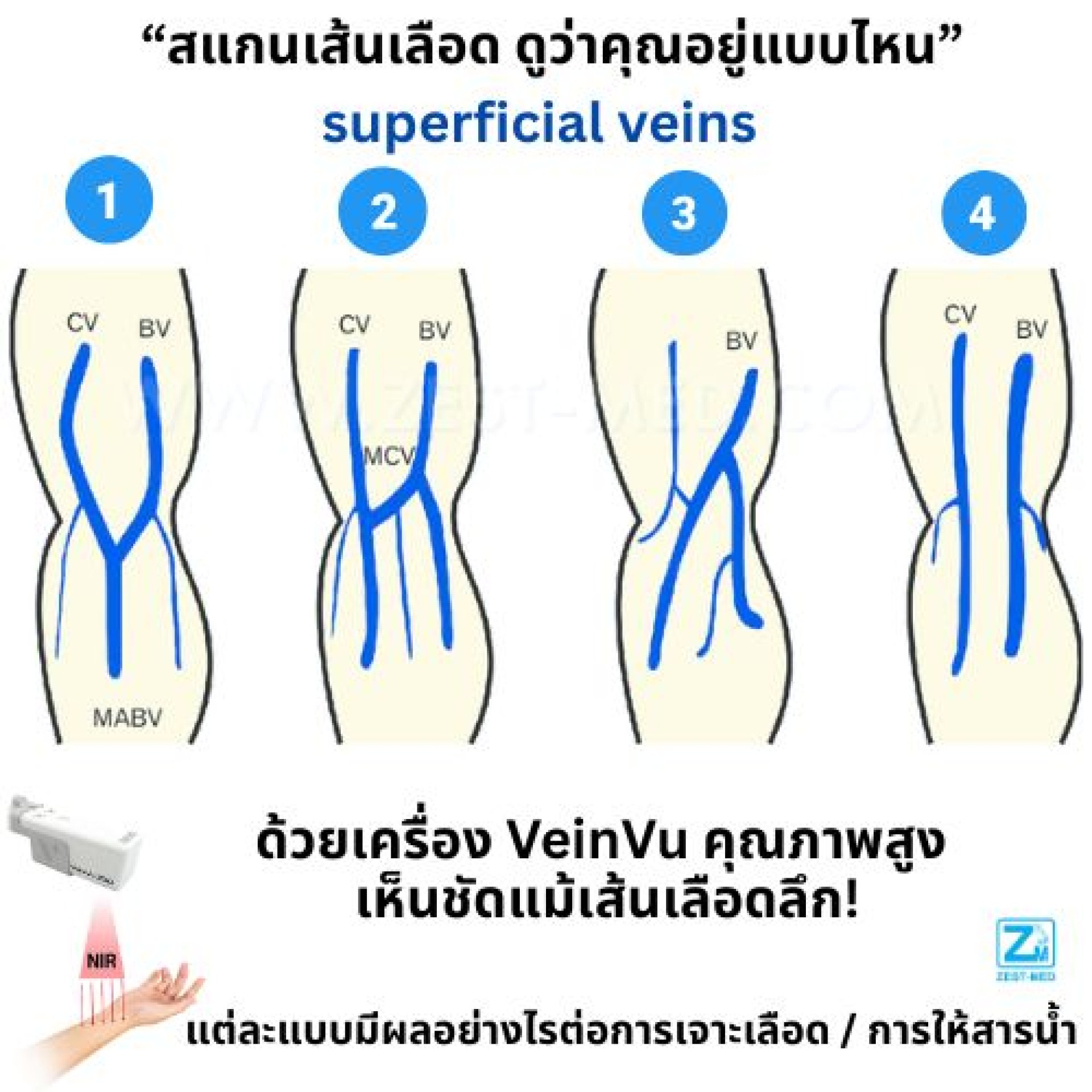
From research to accuracy: 4 patterns of antecubital veins for safe and precise venipuncture
Venipuncture and IV fluid administration are basic procedures encountered daily in medical practice. However, the success of these procedures does not depend solely on the practitioner's skill — the vein structure pattern in the antecubital area also plays a significant role.
Research from Keimyung University School of Medicine, Korea used Infra-red Vein Imaging technology to examine superficial veins in over 200 real subjects, finding 4 main patterns that differ from one another, each of which directly affects the ease or difficulty of venipuncture and IV fluid administration.
4 Patterns of Antecubital Veins
1️⃣ Type I M-Type (46.7%)
Characteristics: Median antebrachial vein is prominent, connecting the Cephalic and Basilic veins
Effect on blood draw: Easy to puncture, clear central vein, reduces risk of repeated puncture
Effect on IV fluid administration: Good flow, suitable for large-volume infusion requiring continuity
2️⃣ Type II H-Type (50.1%)
Characteristics: Median cubital vein directly connects the Cephalic and Basilic veins
Effect on blood draw: Standard position, high success rate, suitable for practitioners of all skill levels
Effect on IV fluid administration: Suitable for long-duration fluid or medication administration at a constant rate
3️⃣ Type III Underdeveloped Cephalic vein (2.0%)
Characteristics: Lateral CV is small or absent
Effect on blood draw: Difficult to puncture, must search for another vein or use advanced techniques
Effect on IV fluid administration: Limited flow, may need to choose another site such as the forearm or back of the hand
4️⃣ Type IV Parallel Type (1.1%)
Characteristics: Cephalic and Basilic veins run parallel, with no central connecting vein
Effect on blood draw: Must choose one side, resulting in positional limitations
Effect on IV fluid administration: Volume and speed depend on the side chosen, suitable for administering smaller amounts of fluid
Why knowing the vein pattern matters
Increases accuracy: choosing the appropriate site reduces the risk of missed punctures
Increases safety: reduces the chance of hematoma and tissue injury
Enables personalized care: choosing techniques and equipment suited to each patient
Educational benefit: used to teach medical students and new staff to become familiar with the diversity of actual anatomical structures
Technology that makes veins visible from the start — #VeinVu200 from Korea
Assessing vein patterns becomes much easier with technology that provides real-time vein visualization. VeinVu 200 uses Advanced Infra-red Vein Imaging technology that can display both superficial and deep veins, even in patients with small or deep veins.
Key features:
Certified by KFDA (Korea Food & Drug Administration)
Sharp real-time imaging helps confirm the position before needle insertion
Lightweight and portable, suitable for use in both clinics and hospitals
Reduces repeated punctures, increases patient comfort, and improves work efficiency
Reference:
Lee H, et al. (2015). Variations of the cubital superficial vein investigated by using the intravenous illuminator. Anatomy & Cell Biology, 48(1), 62–65. DOI: 10.5115/acb.2015.48.1.62
Venipuncture and IV fluid administration are basic procedures encountered daily in medical practice. However, the success of these procedures does not depend solely on the practitioner's skill — the vein structure pattern in the antecubital area also plays a significant role.
Research from Keimyung University School of Medicine, Korea used Infra-red Vein Imaging technology to examine superficial veins in over 200 real subjects, finding 4 main patterns that differ from one another, each of which directly affects the ease or difficulty of venipuncture and IV fluid administration.
4 Patterns of Antecubital Veins
1️⃣ Type I M-Type (46.7%)
Characteristics: Median antebrachial vein is prominent, connecting the Cephalic and Basilic veins
Effect on blood draw: Easy to puncture, clear central vein, reduces risk of repeated puncture
Effect on IV fluid administration: Good flow, suitable for large-volume infusion requiring continuity
2️⃣ Type II H-Type (50.1%)
Characteristics: Median cubital vein directly connects the Cephalic and Basilic veins
Effect on blood draw: Standard position, high success rate, suitable for practitioners of all skill levels
Effect on IV fluid administration: Suitable for long-duration fluid or medication administration at a constant rate
3️⃣ Type III Underdeveloped Cephalic vein (2.0%)
Characteristics: Lateral CV is small or absent
Effect on blood draw: Difficult to puncture, must search for another vein or use advanced techniques
Effect on IV fluid administration: Limited flow, may need to choose another site such as the forearm or back of the hand
4️⃣ Type IV Parallel Type (1.1%)
Characteristics: Cephalic and Basilic veins run parallel, with no central connecting vein
Effect on blood draw: Must choose one side, resulting in positional limitations
Effect on IV fluid administration: Volume and speed depend on the side chosen, suitable for administering smaller amounts of fluid
Why knowing the vein pattern matters
Increases accuracy: choosing the appropriate site reduces the risk of missed punctures
Increases safety: reduces the chance of hematoma and tissue injury
Enables personalized care: choosing techniques and equipment suited to each patient
Educational benefit: used to teach medical students and new staff to become familiar with the diversity of actual anatomical structures
Technology that makes veins visible from the start — #VeinVu200 from Korea
Assessing vein patterns becomes much easier with technology that provides real-time vein visualization. VeinVu 200 uses Advanced Infra-red Vein Imaging technology that can display both superficial and deep veins, even in patients with small or deep veins.
Key features:
Certified by KFDA (Korea Food & Drug Administration)
Sharp real-time imaging helps confirm the position before needle insertion
Lightweight and portable, suitable for use in both clinics and hospitals
Reduces repeated punctures, increases patient comfort, and improves work efficiency
Reference:
Lee H, et al. (2015). Variations of the cubital superficial vein investigated by using the intravenous illuminator. Anatomy & Cell Biology, 48(1), 62–65. DOI: 10.5115/acb.2015.48.1.62


Related Content

Know Your Cubital Superficial Vein Type Elevating safety standards in venipuncture and IV fluid administration — improving puncture success rates and reducing complications.
11 Aug 2025